Embryogenesis
This article, aims to explain the basic levels of
embryogenesis and create the foundation for understanding the concept of EMT
which will be analyzed in another paper.
Everything
begins with a zygote. The zygote is made
out of the zona pellucida, an outer glycoprotein coat , a plasma membrane
(inside of the zona pellucida) and two genetic materials, one of each parent.
The zygote starts dividing into multiple cells, basically cutting into two
cells, which is a process called cleavage. Over time, more and more dividing occurs
, resulting in the creation of many more cells. This is called a morula. (It’s a Greek
word that means mulberry because it somehow looks like it) . Those cells, which
are called stem cells without any differentiation , aren’t very adherent with
each other but they are all very similar. During the next step of this
procedure, the cells begin to get more and more closer together and start to
differentiate, which produces two populations of cells. The one population is called trophoblasts and the
second one embryoblasts. Embryoblasts are the cells on the inside while trophoblasts
are formed on the outside layer.
 |
| https://fertilitypedia.org/edu/therapies/laser-assisted-zona-thinning |
The embryoblasts start to cluster all together in the middle forming a mass called inner cell mass. This actually leaves an empty “bursa” which is called blastocoel.

This whole structure now is called a blastocyst. At this point, the zona pellucida starts to fall apart until it is gone completely while the blastocyst remains. Another bursa forms in the inner cell mass while those cells start to differentiate even more resulting in different type of cells in two layers, the hypoblasts and the epiblasts.
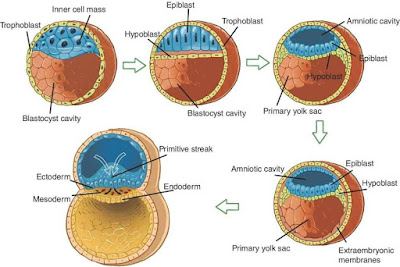
Hypoblasts and epiblasts form a structure of two
layers which is called the bilaminar disk. On the edge of the bilaminar disk, another
formation is created and that is called primitive streak. The primitive streak is the place where the cells begin to migrate.

This causes a change in the structure of the disk. Τhree layers
(germ layers) are formed and that is called a trilaminar disk. The first layer
is called ectoderm, the second is called mesoderm and the third one, endoderm.

Το summarize the whole process until this point, everything started of
with a bunch of cells that were all the same , existing in the whole area
without them clinging to each other.
While the process started to grow, the cells appear to differentiate and start
bonding with each other. The bonding which developed between them is actually the
adhesion of the epithelium cells.
The cells of the mesoderm start to differentiate even more creating a formation called the notochord ( will later form some part of the intervertebral disks).
This formation, will generate another structure in the upper layer, the ectoderm, called the neural plate. The neural plate will fall down to the mesoderm and will create another formation, known as the neural tube. For this structure to be created, the cells of the ectoderm lose a percentage of their adhesion which they have between each other, so they can “dive” away from the surface.


Lastly, the cells of endoderm , mesoderm and ectoderm are
diverse with different characteristics. The mesoderm cells, will create the
mesenchyme and the connective tissue. Because of that, those cells are very distinctive
from the cells of endoderm and ectoderm. They aren’t very adherent to one
another but more attached with their matrix , they have a better ability of movement and they aren’t polarized the way the cells
of the other two layers are. As a consequence, epithelial cells are developed
from all of the three layers while the connective tissue is built only from the
mesoderm cells.
0 comments