Pictures from tumors in the lab


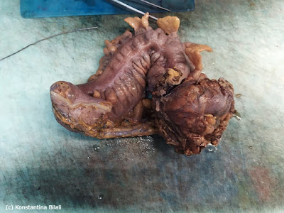
The product is the
result of a left nephrectomy and its size with the perinephric fat is 19x10x8
cm. It is partially covered by the Gerota’s fascia. Also, it connects with the
ureter which is 3 cm long with maximum size of 0,5 cm. After the kidney had
been cut its dimension were 1,5 x 2 cm.
There is an observation of an orange but partially reddish solid and partly
spongy neoplastic tumor, measuring 7x6x4 cm. The tumor is located in middle of
the kidney, 0,1 cm at distance from the
renal pelvis and 1 cm away from the renal capsule. Additionally, the neoplastic
growth reaches closely to the perimetric capsule and to the perimetric renal fat.
The rest of the kidney’s area maintains a medullary architecture and the product
weights 414 kg.

Sigmoid Colon

0 comments